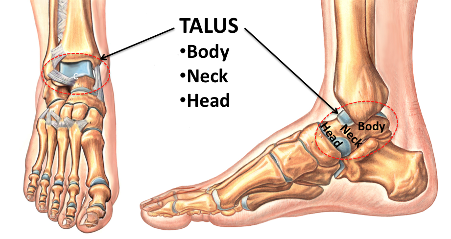
At the time of their article all previous case reports had described sporting avulsion fractures involving small fracture fragments. They believed that in this form of injury, the medial tubercle is likely to be significantly displaced with potential interposition of the flexor hallucis longus tendon. In fact, one group did hypothesize that high energy injuries resulting in fractures of the medial tubercle are very different to sporting avulsion fractures. However, the injury may also occur as a result of direct trauma. This type of avulsion injury was originally described by Cedell in 1974 and most cases have been attributed to this indirect effect. Posterior talar fractures have been most often cited to occur as an avulsion injury secondary to a pronation-dorsiflexion force which causes tension at the insertion of the posterior talotibial ligament. We have reported the successful conservative treatment of a stress fracture of the medial tubercle of the posterior process diagnosed with MR imaging. This is often not the case with conventional anteroposterior, oblique, and lateral radiographs of the ankle and thus surgery has been performed in many cases of this type of injury. They have been most often treated conservatively if diagnosed early. Cases of fractures of the medial tubercle of the talus are very rare in the literature but have typically been cited to occur as a consequence of a pronation-dosiflexion injury.

2).įractures of the posterior process of the talus are potentially very important as the undersurface of this portion of the talus constitutes approximately 25% of the posterior articular facet of the subtalar joint. Routine fast spin echo proton density images further disclosed a discrete low signal intensity fracture line within the medial tubercle without distraction or displacement (Fig. Sagittal fast inversion recovery image demonstrated focal intense bone marrow edema pattern in the far medial tubercle of the posterior process of the talus (Fig. Sequences included: sagittal fast short tau inversion recovery (STIR) (TR/TE 5367/17, FOV 160 mm, ETL 7, slice thickness 3.4 mm, 0 gap, TI 150, matrix 256 × 192, 2 NEX), axial proton density (TR/TE 4917/26, FOV 140 mm, ETL 7, slice thickness 3.5 mm, matrix 516 × 256, 2 NEX), coronal proton density (TR/TE 3650/28, FOV 110 mm, ETL 9, slice thickness 4 mm 0 gap, matrix 512 × 384, 2 NEX), and sagittal proton density (TR/TE 4000/26, FOV 150 mm, ETL 9, slice thickness 3 mm, 0 gap, matrix 512 × 384, 2 NEX). The foot was placed in a neutral position in a quadrature phased array knee coil. MR imaging was performed on a 1.5T MR scanner (General Electric, Milwaukee, WI, USA). There was no obvious injury to bone reported on her radiographs (anteroposterior, lateral, and oblique views) performed at an outside institution so an MRI was ordered to assess the soft tissues and diagnose any subtle injury within the ankle joint.

This discomfort was exacerbated by resisted inversion and a unilateral heel raise. It was sharp in character and at its worst 8 out of 10 on the visual analogue scale but typically 4 to 5 out of 10. On presentation to the clinic at 4-weeks post-injury, she described medial ankle pain deep to the medial malleolus and along the course of the posterior tibial tendon. At 3 weeks she was still feeling discomfort within the ankle, in particular when carrying her twin sons. She rested, iced, and elevated the affected side and used oral anti-inflammatories as needed for approximately 3 weeks. The patient is a 36-year-old female who “missed a step” while descending the stairs in her home, impacted heavily on her right ankle and sustained a supination injury.


 0 kommentar(er)
